In this topic, there are a lot of different concepts that are necessary for understanding the results, I will explain them first.
- Ommatidia: Visual units in insects where the photoreceptors are located.
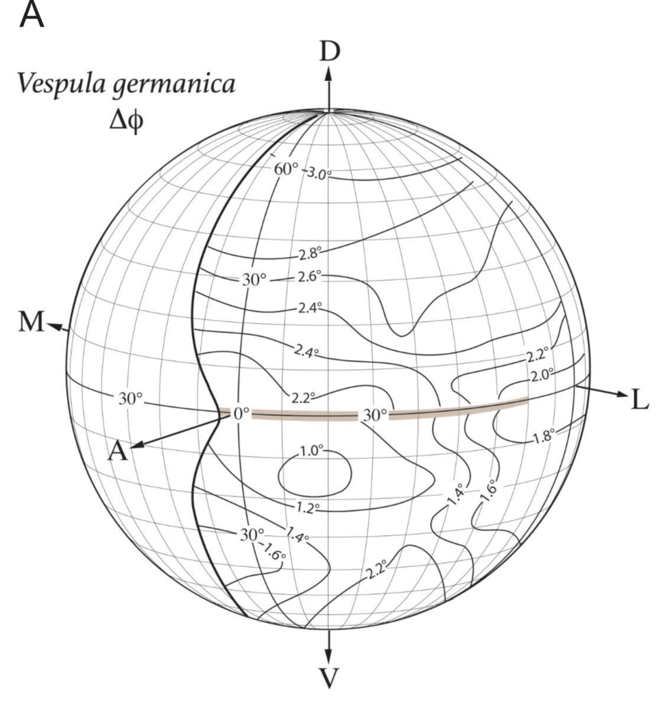
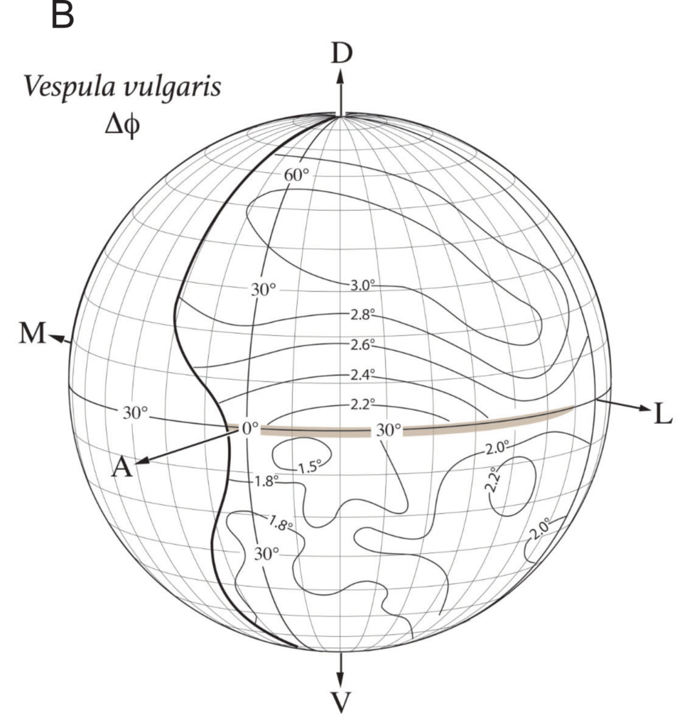
- Interommatidial angle (ΔΦ): The angle between neighbouring ommatidia, the smaller the angle the greater the distance at which objects can be seen.
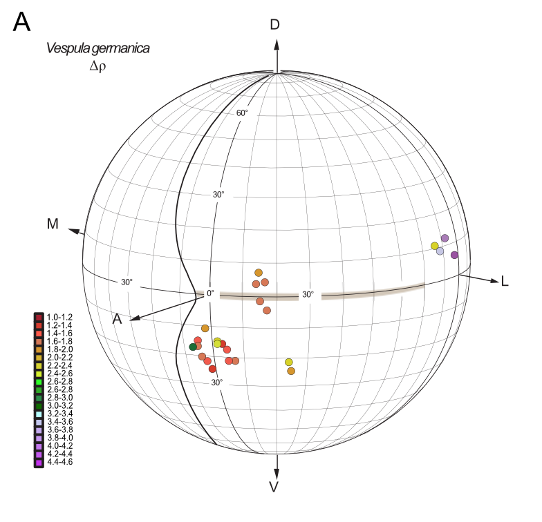
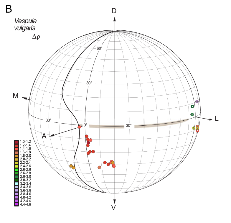
- Acceptance angles (ΔΡ): Angle of the “entrance” of the light and image perception in the ommatidia. The smaller the angle, the higher quality of the optical image.
Both V. germanica and V. vulgaris have well-formed compound eyes and three ocelli on the dorsal surface of the head. They also both possess a cuticular “peninsula” that partially divides the eye into a dorsal and ventral part. It was surmised that this cuticular indentation might create a “blind spot” in the frontal visual field, however as you will see below, this was not the case.
In a compound eye, the packing density of ommatidia is represented by the angle between adjacent ommatidia. This is also known as the interommatidial angle Δφ, which determines the anatomical spatial resolution of the compound eye: the smaller Δφ, the greater the potential resolution. I found that the local Δφ (averaged interommatidial angle) was the smallest in the frontal-ventral part of the eye in both wasps, around 1.0° for V. germanica between -10 and -20 degrees in latitude, and around 1.5° for V. vulgaris between 0 and -10 degrees in latitude (Fig. 2). Thus, both wasps possess an “acute zone”, a region of the eye (and thus visual field) having the greatest spatial resolution. In both wasps, the acute zone is positioned in the frontal eye, just below the horizontal equator. Beneath this acute zone, Δφ increased smoothly towards the most ventral part of the eye.
Although I attempted to sample cells from a range of latitudes and longitudes, the overall variability in both recording location and receptive field width (i.e. acceptance angle Δρ) was too large to allow me to directly generate a complete map of acuity at either the frontal or lateral location. Notably, I obtained several individual cells from the frontal eye region that could be held long enough to scan the receptive field several times, thereby obtaining high-quality receptive field maps, with Δρ below 1.3° degrees (Fig. 1). A comparison of these values in both wasp species reveals that they are very similar: the sharpest values of Δρ are found frontally and the coarser values are found laterally (Fig. 1).
You will find four (4) globes now. Each globe represents the left eye of the wasp that was studied. The thicker vertical line shows the edge of the eye in the head of the wasp. On the left side of this line is the head of the wasp and on the right side is the eye.