Three different methods were employed in this project:
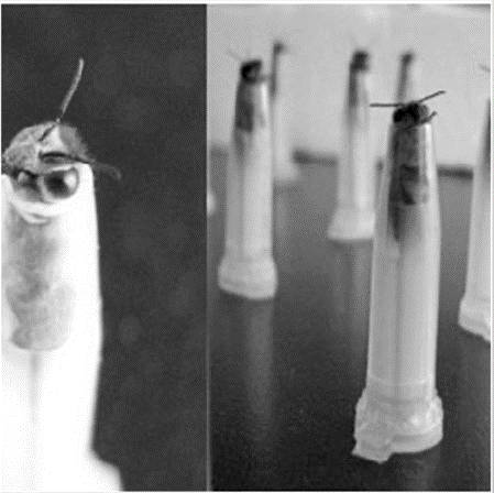
- A single wasp was mounted onto a holder and a triangular window was cut in the dorsal margin on the cornea of the left eye in order to insert an electrode and permit intracellular recordings within the wasp’s left eye (Figure 1).
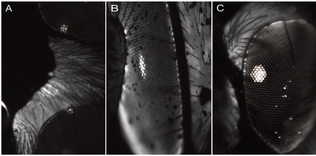
- One wasp was injected with fluorescence dye (Lucifer Yellow which allows the visualisation of the luminous pseudopupils, a small region of visual units of the eye that are directed towards the viewer (Figure 2).
- Wasps’ eyes were removed and transferred into fixatives and solutions for preparation for light, transmission and scanning electron microscopy.